FREE! Click here to Join FunTrivia. Thousands of games, quizzes, and lots more!
Quiz about Parts of the Human Eye

Parts of the Human Eye Trivia Quiz


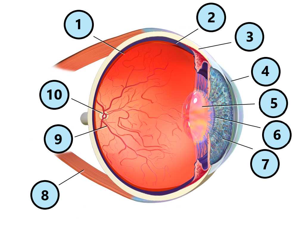
A most complex organ, the human eye requires several different parts in order for it to work properly. Can you label them using this Blausen Medical diagram?

A label quiz by trident. Estimated time: 3 mins.
  1. Home
  2. »
  3. Quizzes
  4. »
  5. Science Trivia
  6. »
  7. Nervous System and the Senses
  8. »
  9. Eyes and Vision

Author
trident
Time
3 mins
Type
Label Quiz
Quiz #
423,696
Updated
Apr 02 26
# Qns
10
Difficulty
Average
Avg Score
7 / 10
Plays
31
Last 3 plays: Guest 72 (2/10), miner8265 (6/10), Guest 98 (10/10).
Click the image to enlarge and get a closer look.
Click on image to zoom
retina lens choroid sclera muscle optic disc iris cornea blood vessels pupil
* Drag / drop or click on the choices above to move them to the answer list.
View Image Attributions for This Quiz
1.   
2.   
3.   
4.   
5.   
6.   
7.   
8.   
9.   
10.   

Most Recent Scores
Today : Guest 72: 2/10
Today : miner8265: 6/10
Today : Guest 98: 10/10
Today : debbitts: 8/10
Today : Guest 207: 6/10
Today : Jaydel: 10/10
Today : Iva9Brain: 7/10
Today : invinoveritas: 4/10
Today : lethisen250582: 10/10

Quiz Answer Key and Fun Facts
1. retina

The retina is a thin layer of tissue at the back of the human eye. It contains specialized cells called photoreceptors (which include rods and cones) that detect light and convert it into electrical signals. These signals travel through the optic nerve to the brain, where they are interpreted as images.

It also includes a small central area called the macula, which allows for the detailed vision needed for reading and recognizing faces.
2. choroid

The choroid is the layer of the human eye located between the retina and the sclera. It is filled with blood vessels that supply oxygen and nutrients to the retina. It also contains dark pigment that absorbs excess light, which prevents light from scattering inside the eye. This feature helps improve the clarity of the images we see.
3. sclera

The sclera is the tough, white outer layer of the human eye. Its main function is to help maintain the eye's shape and protect its inner structures. It is composed of strong connective tissue and acts like a protective shell around the more delicate parts of the eye. It also serves as a surface for muscle attachment, which enables the eye to move in different directions.
4. cornea

The cornea helps protect the eye, while also focusing light. As light enters the eye, the cornea bends it so that it can be properly directed toward the retina. This makes it essential for forming clear images. Unlike most other parts of the eye, the cornea does not contain blood vessels; instead, it receives oxygen directly from the air and nutrients from surrounding fluids.
5. lens

The lens is a flexible structure located behind the iris in the human eye. By changing its shape, the lens adjusts focus so that objects at different distances can be seen clearly, a process called accommodation. This ability allows the eye to switch between viewing things up close and far away.
6. pupil

The pupil is the small, dark opening in the center of the human eye that controls how much light enters. It appears black because the light that enters the eye is absorbed rather than reflected back out. The pupil changes size based on the level of light in the environment, expanding in dim light to allow more light in and contracting in bright light to limit it.
7. iris

The iris is the colored part of the human eye. It surrounds the pupil and controls how much light enters. The iris allows the pupil to change size, as it contains special muscles that make the pupil larger or smaller. Melanin is largely responsible for the coloring of the iris; people with more of this pigment typically have brown eyes, while those with less may have blue or green eyes.
8. muscle

Six muscles, called the extraocular muscles, are attached to the outside of each human eye and work together to move the eye up, down, side to side, and at angles. These muscles allow both eyes to stay aligned and track moving objects smoothly. Smaller muscles inside the eye also help control the size of the pupil and adjust the shape of the lens for focusing.
9. blood vessels

Blood vessels supply oxygen and nutrients to the eye and remove waste products. They are especially important in areas like the retina and the choroid, where cells need a constant supply of energy to function properly.
10. optic disc

The optic disc is a small area at the back of the human eye where the optic nerve connects to the retina. It is the point where nerve fibers leave the eye to carry visual information to the brain. This area does not contain photoreceptors, which means it cannot detect light and creates what is known as a blind spot in vision. Normally, this blind spot is not noticeable because the brain fills in the missing information using input from the other eye.
Source: Author trident

This quiz was reviewed by FunTrivia editor rossian before going online.
Any errors found in FunTrivia content are routinely corrected through our feedback system.
Related Quizzes
1. A Journey into the Eye Tough
2. Doctor, Eye Have a Problem! Easier
3. For Your Eyes Only Average
4. I Can See Clearly Now Average
5. Me, Myself, and Eye Difficult
6. One Minute in Ophthalmology Average
7. Our Eyes Average
8. She Blinded Me with Science Average
9. Sight Unseen - Spectacles Easier
10. Starry Eyed Easier
11. The Cataract Quiz Average
12. The Eyes Have It Average

4/2/2026, Copyright 2026 FunTrivia, Inc. - Report an Error / Contact Us