FREE! Click here to Join FunTrivia. Thousands of games, quizzes, and lots more!
Quiz about Where Am I in the Body

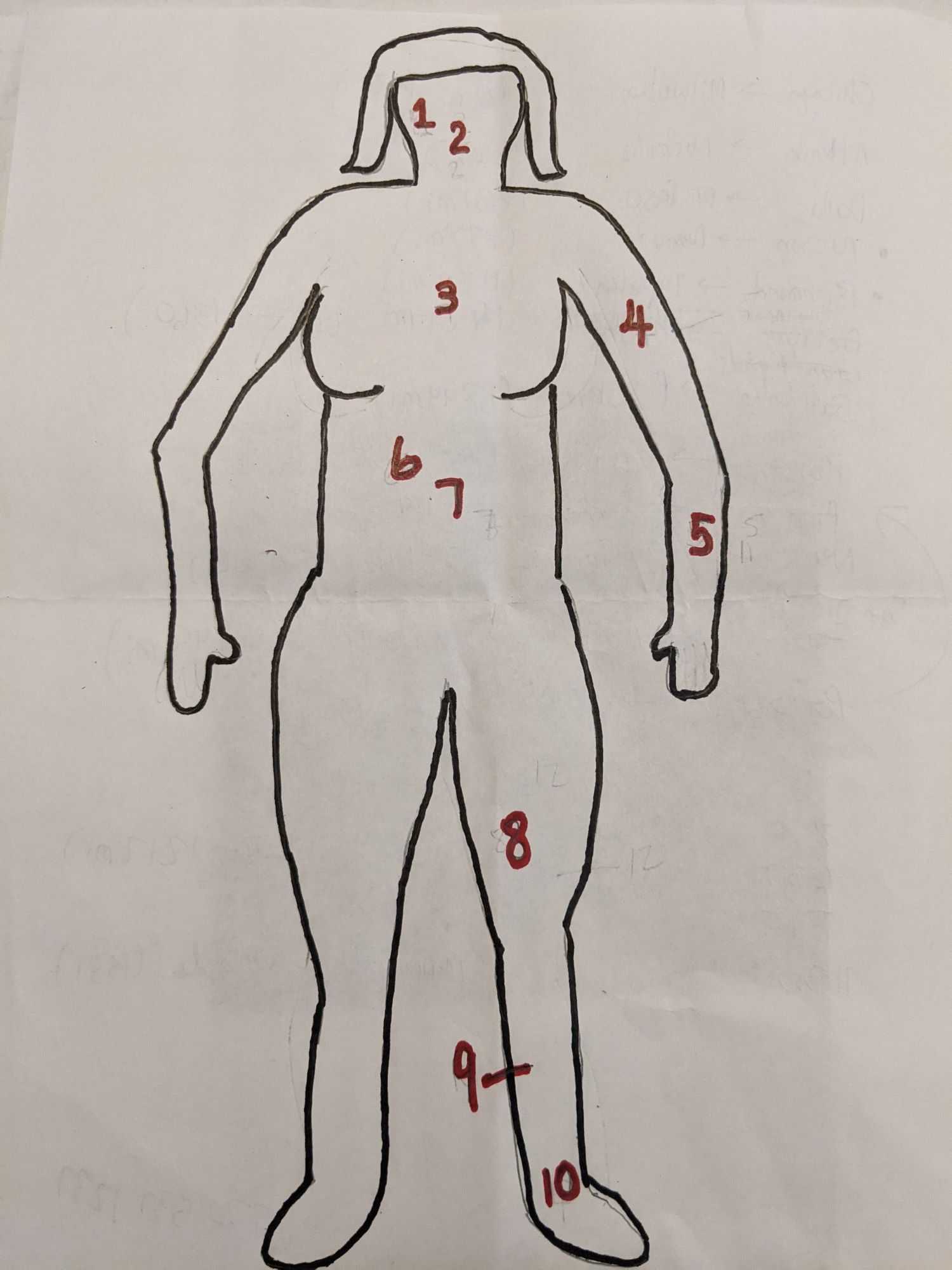
Where Am I in the Body? Trivia Quiz


I am not the only one who enjoyed playing rossian's quiz on "Labelling the Human Body". So, inspired, I have come up with ten more body parts to label for you all to enjoy. Maybe you will find as much humor in my drawing skills as my family did! Enjoy!

A label quiz by BigTriviaDawg. Estimated time: 3 mins.
  1. Home
  2. »
  3. Quizzes
  4. »
  5. Science Trivia
  6. »
  7. Health and Human Biology
  8. »
  9. Human Body

Time
3 mins
Type
Label Quiz
Quiz #
412,921
Updated
Jun 15 23
# Qns
10
Difficulty
Average
Avg Score
7 / 10
Plays
1273
Last 3 plays: Guest 136 (4/10), Guest 73 (5/10), J0key (1/10).
Extensor digitorum Soleus Vastus medialis Humerus Gallbladder Medulla oblongata Sternum Zygomatic Talus Pancreas
* Drag / drop or click on the choices above to move them to the answer list.
1.   
2.   
3.   
4.   
5.   
6.   
7.   
8.   
9.   
10.   

Most Recent Scores
Apr 26 2024 : Guest 136: 4/10
Apr 25 2024 : Guest 73: 5/10
Apr 25 2024 : J0key: 1/10
Apr 25 2024 : Guest 63: 6/10
Apr 25 2024 : Guest 74: 8/10
Apr 25 2024 : Regreg: 8/10
Apr 24 2024 : DRomeo1: 10/10
Apr 24 2024 : Guest 124: 3/10
Apr 24 2024 : Guest 195: 7/10

Quiz Answer Key and Fun Facts
1. Zygomatic

The zygomatic bone is on the side of the face and forms part of the cheek and outer eye socket. The zygomaticus major muscle connects from the zygomatic bone to the corner of the mouth and is the muscle that helps you smile. The zygomatic bone also protects the zygomatic branches of the facial nerves that allow the orbicularis oculi muscle to close the eyelid. Since the zygomatic offers structure to the face, a break or fracture can cause a depression below the eye requiring corrective surgery.
2. Medulla oblongata

The medulla oblongata is a critical part of the brain where it connects to the spinal cord. A nerve highway passes from the brain to the spinal cord through the medulla. Also, critical functions like breathing, heartbeat, and blood pressure are controlled by nerves within the medulla. Like the rest of the brainstem, the medulla is an ancient part of the brain and is sometimes called the "reptilian brain". Damage to the medulla can be very serious including paralysis, loss of sensation, or even respiratory failure.
3. Sternum

The sternum is shaped like a short necktie right under where a necktie would go. The sternum is a bone that protects the lungs and heart and serves as a connection point for the ribs. The costal cartilages connect each rib to the sternum allowing the rib cage to be strong and complete. If a person needs CPR to save their life, the sternum is often fractured or broken in the process.

A broken sternum is likely to cause sharp pain with inhalation or movement. Like a broken rib, sternum injuries usually will heal themselves over the course of a few months.
4. Humerus

The humerus is one of the longest bones in the body and is located in the upper arm. The upper portion of the humerus is rounded and rests in the rotator cuff of the shoulder. Four muscles keep the humerus firmly in the socket. The lower part of the humerus forms a synovial hinge joint (aka the elbow) with the ulna and radius.

The joint allows movement like a hinge, but of course, some twisting can also happen. A broken humerus is a rite of passage for many kids resulting in an itchy arm cast.
5. Extensor digitorum

The extensor digitorum muscle allows a person to extend their digits (fingers). If the hand is resting palm down on a table, the ability to raise the fingers off the table while leaving the arm on the table is the work of the extensor digitorum. This muscle has its proximal connection with the humerus and thus allows it to also move the elbow. Damage to the extensor tendon associated with the muscle results in difficulty in opening the fingers of the hand making it very difficult to use.
6. Gallbladder

The gallbladder is a pear-shaped sack that stores bile made by the liver. The bile helps the body digest fats in a meal and gets released into the duodenum of the small intestines via the bile duct.

Gallstones can develop if the cholesterol and bile in the gallbladder start to harden. The stones can range in size from a small grain of sand to the size of a golf ball. It is possible to have gallstones and not know it, but many people will experience lower abdominal pain. There is a strong correlation between having high cholesterol and developing gallstones. Complications with gallstones usually lead to a cholecystectomy which is the surgical removal of the gallbladder. If the gallbladder is removed, bile can still make its way to the duodenum, it just does not wait for a meal before being released. Complications after surgery include challenges with digesting fatty foods.
7. Pancreas

The two main functions of the pancreas are to release exocrine secretions to help digest sugar, protein, and fat and to release endocrine secretions like glucose and glucagon. The bile duct is used to release all of the exocrine secretions into the duodenum along with sodium bicarbonate to reduce acidity as the material moves from the stomach into the small intestine. Endocrine secretions, like glucose, are released into the bloodstream. Complications of the pancreas can be life-threatening since it serves such an important role in food digestion and blood sugar regulation. Also, cancer of the pancreas is one of the most aggressive and deadly forms of cancer often with little hope for remission.
8. Vastus medialis

The vastus medialis is one of the four muscles that make up the quadriceps of the thigh. The muscle is on the interior of the quadriceps and travels the entire length of the femur to attach to the quadriceps tendon. On the distal end, the vastus medialis connects to the patella and helps keep the knee cap in place. Knee pain is frequently attributed to issues with the vastus medialis. Proper physical therapy can help keep the muscle healthy and alleviate the knee pain it may have caused.
9. Soleus

The soleus muscle is at the back of the leg under the gastrocnemius (calf) muscle. The soleus connects to the back of the shin and the heel via the Achilles tendon. The soleus is critical for walking, running, and jumping. Commonly pain a new runner might consider being shin splints is actually strain to the soleus muscle. Since the gastrocnemius muscle is much stronger, it is able to handle more strain than the soleus can. Slowly increasing the intensity of exercise is critical to allow the soleus to grow, get stronger, and provide the support needed for the athlete.
10. Talus

The talus is more commonly known as the ankle bone. Both the fibula and tibia rest on top of the talus and the talus rests on top of the heel bone. The talus is one of the bones that allow articulation of the foot. It also allows pressure and weight to be transferred across the foot and thus contributes to the stability of the lower body.

While the talus is a strong bone, if it gets fractured it can take a very long time to heal since it has very limited blood flow. An injury here may require several months to a half year or longer in a boot to keep the foot immobilized.
Source: Author BigTriviaDawg

This quiz was reviewed by FunTrivia editor rossian before going online.
Any errors found in FunTrivia content are routinely corrected through our feedback system.
4/27/2024, Copyright 2024 FunTrivia, Inc. - Report an Error / Contact Us