
Where Does the Blood Go? Trivia Quiz
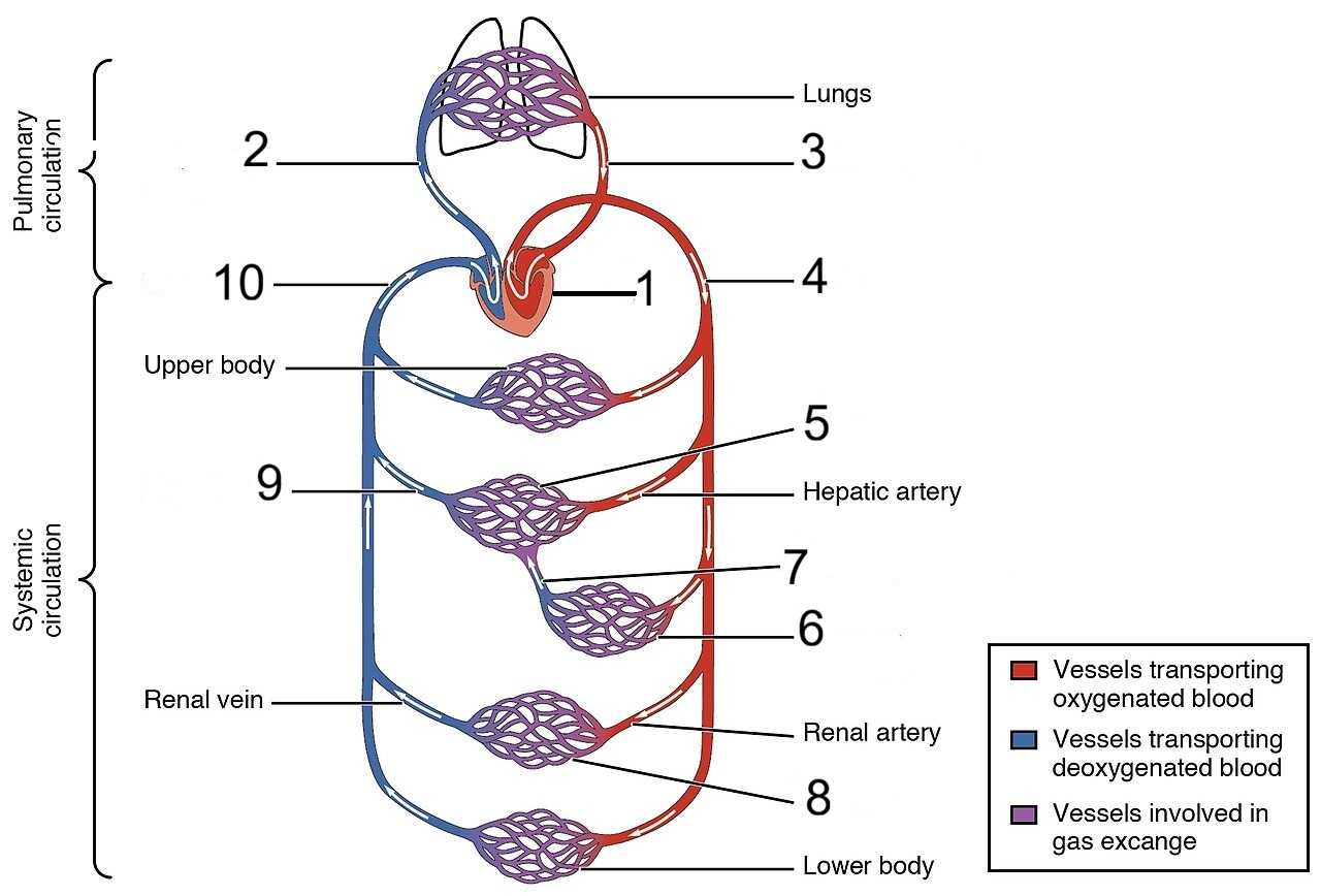
Some of the labels have fallen off this schematic diagram of the human circulatory system. Can you put them all back in the correct spot?
A label quiz
by looney_tunes.
Estimated time: 3 mins.
| 1. |
| 2. |
| 3. |
| 4. |
| 5. |
| 6. |
| 7. |
| 8. |
| 9. |
| 10. |